Ear Anatomy Explorer
Click any labeled structure in the outer, middle, or inner ear to learn what it is, how it works, and exactly how microtia affects it.

If your child was diagnosed with microtia: Understanding which structures are affected explains the hearing impact and what reconstruction involves. Dr. Bonilla reviews each child's specific anatomy at consultation.
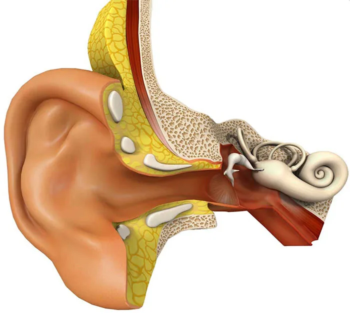
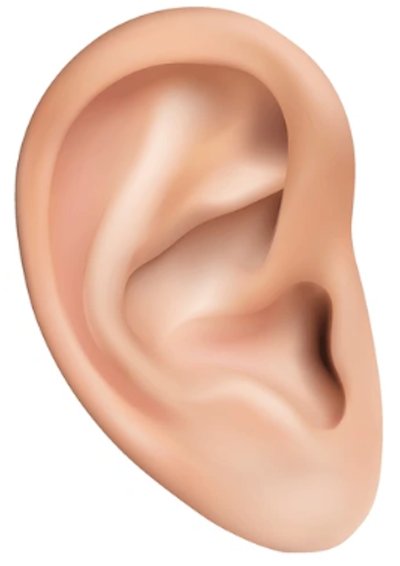
Outer Ear Anatomy
The outer ear includes the visible auricle and the opening of the ear canal and plays an important role in overall hearing function. Its main job is to collect sound waves and direct them inward toward the eardrum. Structures such as the helix, antihelix, tragus, concha, and lobule give the ear its shape and also help with sound localization.
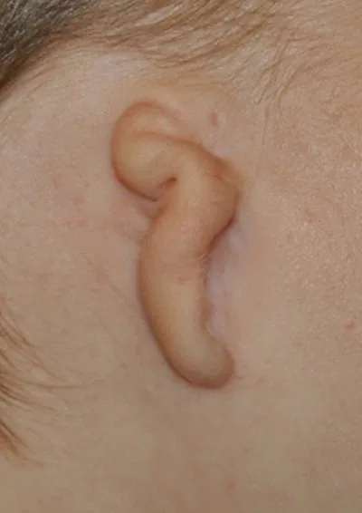
In microtia, the outer ear is underdeveloped to varying degrees depending on the grade. This affects appearance, but it also matters because the outer ear normally helps funnel sound efficiently toward the middle ear. Understanding the anatomy of the auricle helps families better understand how microtia is classified and why reconstruction planning varies from child to child.
Tap a region to highlight it. Tap again to learn more.
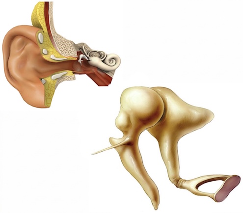
Middle Ear Anatomy
The middle ear begins at the tympanic membrane, or eardrum, and includes the three small hearing bones: the malleus, incus, and stapes. These structures vibrate in response to sound and amplify that energy before passing it into the inner ear, and plays a critical role in how sound is transmitted during the hearing process. The Eustachian tube also plays an important role by helping equalize pressure behind the eardrum.
In children with microtia and aural atresia, the middle ear may also be affected along with the outer ear. The ear canal may be narrow or absent, preventing sound from reaching the eardrum and ossicles efficiently. Even when this pathway is interrupted, the inner ear is usually still capable of processing sound normally, which is why hearing intervention can be so effective.
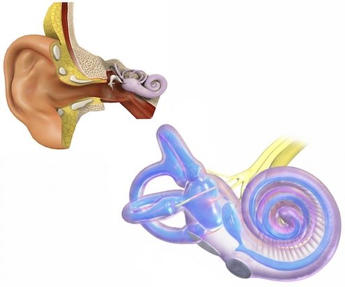
Inner Ear Anatomy
The inner ear contains the cochlea, which converts sound vibrations into electrical signals for the hearing nerve, and the vestibular system, which helps maintain balance, and is essential for both hearing and balance. Key structures include the cochlea, semicircular canals, vestibule, and vestibulocochlear nerve. Unlike the outer and middle ear, these structures are responsible for actually processing sound and sending it to the brain.
In most children with microtia, the inner ear develops normally. This is one of the most important concepts for families to understand, because it explains why many children with microtia are not deaf even when the outer ear looks significantly different. Hearing loss is usually conductive, meaning the hearing organ itself is present and healthy but the pathway that delivers sound is disrupted.
How Ear Anatomy Relates to Microtia and Hearing
Microtia affects the external ear and is frequently associated with aural atresia, which can block sound from reaching the eardrum and middle ear bones. The degree of outer ear difference does not always predict hearing ability exactly, which is why formal hearing testing is so important. By understanding how the outer, middle, and inner ear normally work together, families can better understand both the appearance-related and hearing-related aspects of microtia.
This page is designed to help parents visualize that relationship clearly: the outer ear collects sound, the middle ear transmits it, and the inner ear converts it into signals the brain can understand. In most microtia patients, the inner ear remains intact, which is why bone-conduction hearing solutions can make such a significant difference.
Dr. Bonilla Explains Ear Anatomy in His Own Words
Understanding ear anatomy is the foundation of understanding microtia. Dr. Bonilla walks through all three regions — outer, middle, and inner ear — in plain language designed for families, explaining what is absent in microtia and why the inner ear being normal is such genuinely good news.
If you have questions after watching, Dr. Bonilla's team is happy to help.
Request a Consultation With Dr. BonillaWatch Dr. Bonilla explain the anatomy of the outer, middle, and inner ear and how each structure contributes to hearing.
Questions About Your Child’s Specific Anatomy?
Dr. Bonilla examines each child directly and explains exactly what reconstruction involves for their specific grade and anatomy — outer ear, hearing, and all.