The Four Grades of Microtia — What Each One Means for Your Child
Every grade is treatable. This page explains what each grade looks like, how it affects hearing, and so you can understand your child’s specific situation clearly.

Understanding the Grading System
The four grades describe how much of the outer ear formed during fetal development. They are a classification system, not a prognosis. Grade IV is not “worse” than Grade I in any outcome sense — all four grades produce reliable, well-documented results with rib cartilage reconstruction in experienced hands. The grade shapes the surgical approach; it does not determine the quality of the result. For parents new to the diagnosis, understanding microtia as a condition is a helpful place to start before exploring the grading details below.
One critical point: the grade of the outer ear does not predict hearing. The inner ear — the cochlea and auditory nerve — develops from entirely different embryological structures and is normal in the vast majority of microtia cases regardless of grade. The hearing loss in microtia is almost always a pathway problem, not a nerve problem.
What Grade I looks like
All recognizable anatomical structures are present: the helix (outer rim), antihelix, tragus, concha, and earlobe. The ear is clearly identifiable as an ear from any normal viewing distance. What distinguishes it is that it is smaller than normal, and one or more structural elements may be underdeveloped or misshapen.
Common features: a helix that does not complete its curve, a shallower concha, a smaller or absent tragus, or an overall ear noticeably smaller than the opposite side. In some cases the differences are subtle; in others the asymmetry is obvious to any observer.
How Grade I affects hearing
Grade I typically causes mild to moderate hearing reduction — less than higher grades because more outer ear structure is present. Many children with Grade I hear well enough for normal speech and language development. However, they still benefit from audiology evaluation and monitoring, as even subtle hearing differences between ears can affect spatial hearing and classroom performance.
Dr. Bonilla's surgical approach for Grade I
Grade I patients rarely require surgery. A slightly smaller but naturally formed ear is almost always preferable to a reconstructed one — and in most Grade I cases the ear retains enough natural structure that reconstruction would offer little meaningful improvement. However hearing evaluation is still essential — an Auditory Brainstem Response test should be performed early to confirm the hearing status on the affected side, as hearing can still be affected even when the outer ear appears nearly normal. Dr. Bonilla monitors Grade I patients carefully over time — tracking hearing, development, and the child’s own perspective as they grow. If any intervention is ever considered it is discussed openly with the family and the child when age appropriate.
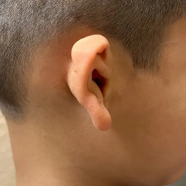
What Grade II microtia looks like
Grade II is defined by a partially formed outer ear — some recognizable structures are present, but a significant portion is absent. The most common presentation is an upper or lower portion of the ear that has formed with some identifiable shape, while the rest is absent or vestigial. A small hook-shaped cartilage remnant with a displaced earlobe is also common. The ear canal is typically absent or severely narrowed in Grade II.
How Grade II affects hearing
With the ear canal typically absent, Grade II produces a moderate to moderately severe conductive hearing loss of approximately 45–60 dB. The inner ear is almost always fully functional — this is a blockage problem, not a nerve problem. Bone conduction typically reveals normal inner ear function. A bone-anchored hearing aid (BAHA) is the standard early hearing management strategy.
Dr. Bonilla’s surgical approach for Grade II
Dr. Bonilla typically builds the full ear framework from rib cartilage in one surgery, avoiding the need for any additional stages.
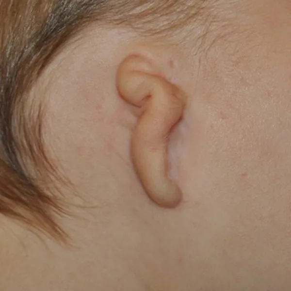
What Grade III microtia looks like
Grade III is what most people picture when they hear the word microtia. It is by far the most common presentation — approximately 7 in 10 children with microtia have Grade III. The defining feature is a small, irregular cartilage remnant — often described as peanut-shaped, sausage-shaped, or “crumpled.” This remnant is residual cartilage tissue that did not develop into a recognizable ear structure. Below or attached to it is a displaced earlobe — a small tag of skin representing surviving lobule tissue. There is no ear canal.
How Grade III affects hearing
With no ear canal, Grade III produces a moderate-to-severe conductive hearing loss of approximately 40–60 dB on the affected side. But the unaffected ear does most of the work. Most children with unilateral Grade III develop normal speech and language with no intervention — the brain compensates remarkably well. There are real-world impacts though: difficulty in noisy environments, inability to locate the direction of sounds, and listening fatigue. Many families choose a BAHA softband during early childhood; others monitor without a device.
Dr. Bonilla’s Surgical Approach for Grade III
Depending on the amount of available skin and its elasticity, Dr. Bonilla may be able to complete Grade III reconstruction in two surgeries instead of three. If there is not enough skin, the standard three-stage protocol is followed as described below.
Stage 1: The peanut remnant is preserved while the rib cartilage framework is harvested from ribs 6, 7, and 8. The framework is hand-sculpted to exactly match the opposite ear in every dimension, then placed in a precisely sized skin pocket. A drain maintains tight skin-to-cartilage contact during healing — this is what creates the anatomical detail visible in the framework during healing.
Stage 2: The preserved earlobe is rotated to its correct anatomical position. The tragus is formed. The conchal bowl is deepened. Because only native skin is used on the visible surface, there is no color difference anywhere on the ear.
Stage 3: The ear is elevated from the head to achieve natural projection. A skin graft placed behind the ear maintains this elevation permanently. BAHA implant can be placed concurrently if desired. The reconstruction is complete.
In some cases — where sufficient skin is available, determined either before or during surgery — Grade III reconstruction can be completed in two stages, finishing within approximately two to three months.
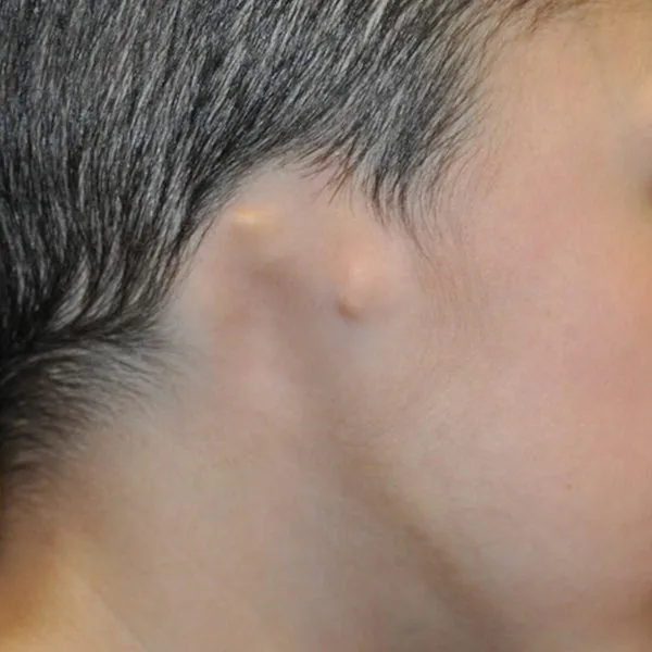
What Grade IV (anotia) looks like
Anotia is the complete absence of the external ear. There is no peanut-shaped remnant, no earlobe, no cartilage nub — the side of the head is smooth where the ear would be. No ear canal, no external opening of any kind. The skin over the area is intact and completely normal; it simply has no ear on it.
Anotia is rare — approximately 5% of microtia cases. It occurs more often in bilateral microtia (both ears affected) than in unilateral cases, though it can and does present on one side alone.
Surgical Considerations for Grade IV
Parents hearing “there’s nothing there at all” naturally feel more alarmed than parents of Grade III children. Anotia cases, while requiring careful planning, benefit from an undisturbed skin surface that allows precise framework placement without navigating misformed tissue. The reconstructed ear is placed exactly where it belongs, on undisturbed skin.
Because no earlobe tissue exists, on the second surgery the framework is elevated and the lower part of the framework is considered to be the earlobe.
Dr. Bonilla’s surgical approach for Grade IV
On the first stage, Dr. Bonilla forms the cartilaginous framework of the full ear. On stage two, because there is no lobule or earlobe, the whole ear is separated and elevated.
Because there is no earlobe present in Grade IV anotia the earlobe transposition stage is not required. Reconstruction follows a modified two-stage approach — Stage 1 establishes the cartilage framework and Stage 2 completes the elevation and final positioning — typically finishing in two surgeries rather than three.
What It Looks Like
Bilateral microtia means both ears are affected. Each ear may present at a different grade — one ear may be Grade III while the other is Grade II, or both may be the same grade. The condition occurs in approximately 10% of microtia cases and presents unique considerations for both hearing and reconstruction that differ meaningfully from unilateral cases.
Hearing Management
When both ears are affected hearing intervention is not optional — it is essential from the earliest possible age. A bone conduction hearing aid softband should be fitted ideally within the first two months of age. Unlike unilateral microtia where the unaffected ear compensates, bilateral microtia means the child has no natural hearing pathway on either side. Without early intervention speech and language development can be significantly affected. An Auditory Brainstem Response test should be performed as early as possible to confirm inner ear function — which is almost always intact — and to establish the foundation for a hearing plan.
Surgical Approach
For Grade III bilateral microtia Dr. Bonilla uses a coordinated staging approach across both ears — combining stages so that the entire bilateral reconstruction is typically completed within approximately six months. In some cases where sufficient skin is available this can be completed even faster. For bilateral Grade II a single stage on each ear may be all that is needed. For bilateral Grade IV the modified two-stage approach applies to each ear, potentially shortening the overall bilateral timeline further. Dr. Bonilla plans each ear individually and stages the surgeries to allow full healing between procedures.
What Families Should Know
Bilateral microtia is a more complex presentation but it is one Dr. Bonilla has extensive experience with. Families with bilateral children are encouraged to reach out early — not because surgery is urgent, but because the hearing plan needs to begin immediately and the surgical roadmap benefits from early planning. Many children with bilateral microtia go on to live full active lives with natural reconstructed ears and well-supported hearing.
How the Grades Compare
One thing is consistent across every row: the outcome quality from rib cartilage reconstruction.
| Grade I | Grade II | Grade III | Grade IV | |
|---|---|---|---|---|
| Frequency | ~5–10% | ~15–20% | ~70% — most common | ~5% |
| Recognizable ear? | Yes — small, some structures | Partial | No — remnant only | No — nothing visible |
| Earlobe present? | Yes | Sometimes | Yes (displaced) | No — created in surgery |
| Ear canal present? | Often — may be stenotic | Typically absent | Absent | Absent |
| Hearing loss | Mild–moderate (~25–40 dB) | Moderate–severe (~40–60 dB) | Moderate–severe (~40–60 dB) | Moderate–severe (~40–60 dB) |
| Inner ear (cochlea) | Normal | Normal | Normal (~95%) | Typically normal |
| BAHA recommended? | Often not needed | Yes — from infancy | Optional | Yes — urgent if bilateral |
| Canal surgery eligible? | Possible — CT determines | Select cases | Select candidates | Rarely |
| Surgical stages | Dr. Bonilla rarely recommends surgery | One surgery | 1 to 3 surgeries | 2 |
| Surgery timing | Ages 6–9 | Ages 6–9 | Ages 6–9 | Ages 6–9 |
| Result quality | Surgery normally not recommended | Excellent | Excellent | Good — depending on amount of skin |
Related Resources
Understanding the grade is the beginning of the process, not the end. These are the most useful next resources for families who just received a diagnosis.
All Four Grades Are Candidates for Reconstruction
Understanding your child’s specific grade is the foundation for everything that follows — surgical timing, hearing management, and long-term planning. A consultation with Dr. Bonilla provides a personalized assessment of all of these. Dr. Bonilla evaluates each child individually — grade, timing, expected result, hearing options — tailored to your child’s exact anatomy. Telehealth available worldwide.